A Deeper Look into the Brain
People observe and interpret other people’s actions in order to predict future behavior. This is a central skill for everyday life. At the Laboratory for Social and Neural Systems Research, we add to the understanding of human behavior by observing decision-making in experiments and using neuroscience to take a deeper look into people’s brains.
However much data one manages to generate, simply observing behavior, or asking people to explain the thinking behind their actions, will not suffice to fully understand it. This is most visible in the case in which behavior is not in line with one’s own preferences, is inconsistent, or contradicts rationality. Until recently, such contradictions were conveniently put into a black box labeled “subconscious.”
For a scientist, a black box is an unsatisfactory answer. Advances in neuroscience have given us insights into the brain and paved the way for a new area of research: neuroeconomics combines the fields of neuroscience, biology, psychology, and economics. The findings shed light on the subconscious and help us understand the causes of human behaviors such as altruism, egoism, risktaking, or self-control at the neural level.
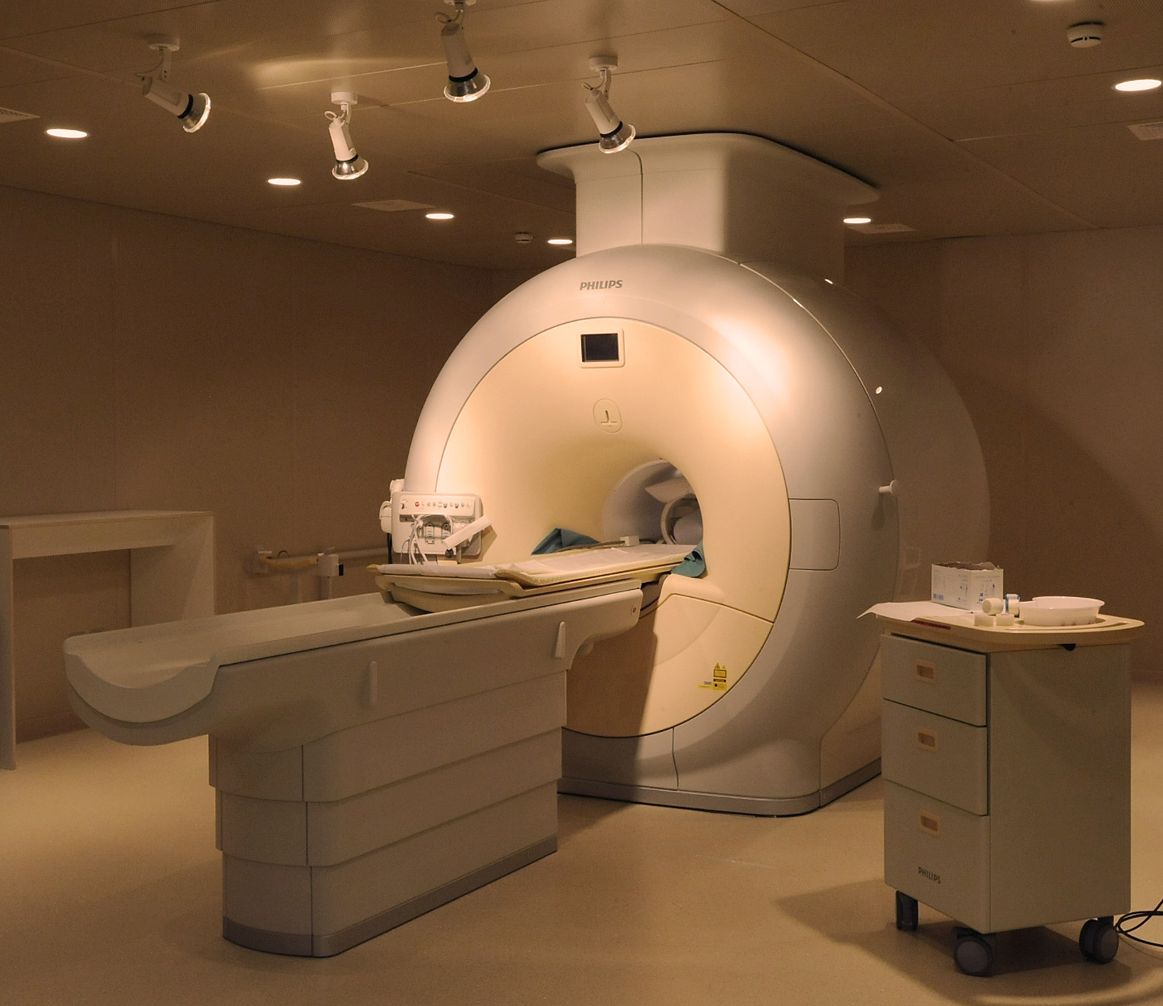
In 2007, thanks to a generous donation by Branco Weiss, the Department had the unique chance to establish the Laboratory for Social and Neural Systems Research (SNS-Lab) in the basement of the University Hospital Zurich. The heart of the lab is an fMRI Scanner, allowing visualization of active brain areas during cognitive processes. In addition, the lab is home to EEG and TMS apparatuses (see box on next page) as well as eye-tracking tools and a behavioral experiment lab. Whereas researchers in other institutions only have access to fMRI scanners when these are not needed for medical patients – which means late evenings and nighttime – our researchers have unlimited access. The scanner runs around 2,000 hours a year, an average of 40 hours per week. Having a dedicated fMRI scanner for research purposes puts the Department in an advantageous position and has led to it becoming a pioneer in the field.
The SNS-Lab shares this privilege by opening the scanner to researchers outside the Department for 20 percent of the time. Currently the University Hospital Zurich and the Psychiatric Hospital of the University of Zurich as well as the Institute for Biomedical Engineering and the Department of Health Sciences and Technology of the ETH use the scanner regularly for research purposes. The research topics are broad: overweight, toothache, and, of course, economic behavior in all its shapes and sizes.On an average day a team is at work to understand impatience and self-control. Based on findings of a previous behavioral experiment, the scientists know the following: if the temporo-parietal junction, the area responsible for mental projections and considerations concerning the future, is disrupted through Transcranial Magnetic Stimulation (TMS), people tend to make more impulsive and selfish decisions. However, this observation alone does little to tell us if the change in behavior is based on a change of self-control mechanisms, i.e. a change in temporal discounting preferences, or a completely different reason. To understand the underlying mechanisms, the original behavioral experiment is repeated in the fMRI scanner in order to identify the activity of the various areas in the brain during the decision-making process with and without the TMS treatment.
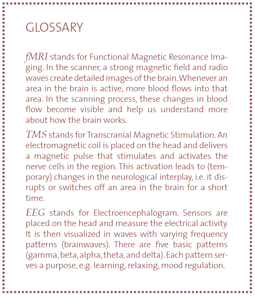
Watching this from the control room through the mirrored window, one gets a sense of the precision, speed, and teamwork necessary. After the test person has gone through the experiment in a first round, he or she is rolled out and a coil generating Transcranial Magnetic Stimulation (see box opposite) is positioned just behind and above his/her ear, precisely where the temporo-parietal junction lies. A crackling sound accompanies the stimulation for about 40 seconds and then he is rolled back into the scanner. The lab manager and two scientists quickly leave the room, close the door and the experiment is repeated.
The scanner generates around 40 slice images of the brain in two seconds. Each of these slices is around 3mm thin. Depending on the research question and the areas of the brain in concern, different settings and scanning speeds are required. A full scan of the brain takes between 1.5 and 3 seconds. Usually, the subject in the scanner is asked to make decisions by pressing yes or no buttons while the scanner is running and taking images of his brain. As the scientists want images of brain activity milliseconds before the subject makes the decision, the timing of the automated questions and scanning have to be aligned very precisely. The Department has a dedicated IT Engineering team for this.
To ensure ongoing precision, a physicist recalibrates and tests the scanner every week. However, there is an imperfection that cannot be corrected so easily: magnetic fields do not have perfectly even structures. These irregularities have an influence on the images the scanner creates and the results need to be recalculated to exclude the distortion created by the inhomogeneity of the magnetic field.
The strong magnetic field generated by the fMRI also means that special rules apply in the lab. Anything magnetic turns into a dangerous weapon within the vicinity of the scanner. Therefore, only people who have ran through an intensive lab security training are allowed to use the machinery, as security is a top priority.